
TL;DR
Stanford researchers developed interferometric Image Scanning Microscopy, or iISM, to image structures moving inside living cells without fluorescent labels. Their paper reports resolution of about 120 nanometers; the team says the technique could complement fluorescence microscopy in studies of cell activity, disease and drug responses.
Researchers at Stanford University have developed a microscope that can show structures moving and interacting inside living cells without fluorescent labels, according to a study published in Light: Science & Applications. The system, called interferometric Image Scanning Microscopy (iISM), achieves resolution of about 120 nanometers, the researchers report, offering a way to track cellular activity while avoiding some limitations of fluorescent imaging.
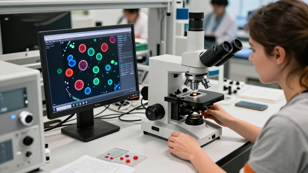
The Stanford team combined interferometric scattering microscopy with camera-based detection adapted from advanced confocal microscopes. In scattering microscopy, a laser illuminates a sample and cellular structures scatter some of its light. Because that scattered signal is weak, a second beam amplifies it through interference so the instrument can detect the structures. The detector array collects multiple views of a region, which the system uses to build a sharper image.
The paper describes iISM as a label-free method with resolution of approximately 120 nanometers. Fluorescent labels are commonly used to make selected cell components visible, but they can fade during observation, can be difficult to introduce and may affect the structures being studied. The researchers say iISM can instead capture multiple cellular structures in motion without adding those markers.
The team also reports that iISM uses substantially less illumination power than comparable label-free methods that produce high-contrast images. Lower exposure may reduce photodamage, in which imaging light harms cells or disrupts normal function. The researchers present the instrument as a way to examine cells over time and study how they respond to drugs or disease-causing organisms, not as a replacement for other microscopy methods.
A New View of Cell Activity
Observing several cell structures in motion without fluorescent labels could help researchers study interactions that are difficult to capture when only a limited number of components can be marked. That may be useful for investigating how cells change in response to drugs or infectious organisms, and for research involving cancer, infectious disease and plant-microorganism interactions, areas the team identifies as potential applications.
The reported resolution and low illumination are relevant because live-cell imaging must balance detail against the effects of observing the sample. Fluorescence microscopy remains valuable when researchers need molecular specificity. The Stanford researchers say iISM could add label-free context and dynamics alongside that specificity, allowing different methods to address different parts of a biological question.
Those applications remain prospective. The report describes an imaging advance, not a new treatment, a clinical test or proof that the system will change patient care. Its immediate significance is that researchers may have another tool for studying cellular behavior under observation conditions intended to be less disruptive.
lab-grade interferometric microscope
As an affiliate, we earn on qualifying purchases.
As an affiliate, we earn on qualifying purchases.
How the Imaging Method Works
Interferometric scattering microscopy detects light scattered by tiny structures in a sample. The Stanford system pairs that weak scattered signal with a second laser beam, using interference to make the signal easier to detect. The method therefore does not depend on fluorescent substances to make structures visible.
The team added an array detector inspired by advanced confocal microscopy. Rather than relying on a single detector behind a pinhole, the camera-based array gathers multiple views of the same region. Combining those views helps produce a more detailed image. The study reports approximately 120-nanometer resolution for this label-free approach; the source material does not provide a comparison dataset or a detailed account of how that figure was measured.
Senior author W.E. Moerner received the 2014 Nobel Prize in Chemistry for work related to super-resolution fluorescence microscopy. The Stanford report describes iISM as complementary to fluorescence imaging. That distinction matters: fluorescence can identify specific molecules, while the new approach is intended to show cellular structures and their motion without requiring labels.
“This new microscope provides a fantastic new view into the cell, where you can see the tiny structures and machines in the cell moving, changing, and interacting without having to add fluorescence to observe them.”
— W.E. Moerner, senior author and Stanford chemistry professor
As an affiliate, we earn on qualifying purchases.
Questions Beyond the Resolution Figure
The available report does not specify how the approximately 120-nanometer resolution was tested, which cell types were imaged, or how long cells were observed in the experiments. It also does not give enough information to compare the system’s performance across different samples or laboratories. Those details are needed to judge how broadly the method can be applied.
The stated uses in cancer research, drug development and infectious-disease studies are potential applications, not demonstrated clinical outcomes. It is not clear from the supplied material how the microscope performs in specific experiments involving drugs or disease-causing organisms, or when other researchers may be able to use the system. The instrument’s practical limits and availability are also not stated.
label-free cellular imaging device
As an affiliate, we earn on qualifying purchases.
As an affiliate, we earn on qualifying purchases.
Testing the Microscope in Research
The next step is for researchers to apply iISM to biological questions where observing multiple structures over time, with limited illumination and no fluorescent labels, could add useful information. The team points to studies of cell responses to drugs and disease-causing organisms as possible directions, as well as work on plant interactions with microorganisms.
Further experiments and independent use will help establish how reliably the method works across cell types, how long it can image cells, and where it offers advantages over existing approaches. The researchers’ stated plan is complementary use with fluorescence microscopy, combining molecular specificity from labels with the label-free view of cellular context and motion.
high resolution biological microscope
As an affiliate, we earn on qualifying purchases.
As an affiliate, we earn on qualifying purchases.
Key Questions
What is Stanford’s new microscope called?
It is called interferometric Image Scanning Microscopy, or iISM. The system combines interferometric scattering microscopy with camera-based detection.
Does iISM use fluorescent labels?
The method is designed to image cellular structures without fluorescent labels. The researchers say it may complement fluorescence microscopy, which can provide molecular specificity.
What resolution did the study report?
The report gives a resolution of approximately 120 nanometers for the label-free approach. The supplied source material does not describe the full measurement protocol or comparison conditions.
Could the microscope help study disease or drugs?
The researchers identify drug responses and encounters with disease-causing organisms as potential areas of study. These are proposed research applications; the report does not establish clinical benefits or outcomes.
Source: rss